Upper Leg Tendon Anatomy : Lameness, Recent Front Limb - Horse Side Vet Guide : The biceps is a muscle on the front part of the upper arm.
byAdmin-
0
Upper Leg Tendon Anatomy : Lameness, Recent Front Limb - Horse Side Vet Guide : The biceps is a muscle on the front part of the upper arm.. Jun 18, 2018 · the upper leg is often called the thigh. The biceps is attached to the arm bones by. During an open surgery, an incision is made in the back of the leg and the achilles tendon is stitched together. The muscle descends medially, condensing into a tendon that runs down the leg, between the gastrocnemius and soleus. The posterior upper leg muscles provide your knees with mobility (extension, flexion and rotation) and strength.they work closely with your quadriceps muscles at the front of your thigh, your gluteal muscles, and your calf muscles to ensure proper movement of your leg and hip.
The biceps is attached to the arm bones by. It is absent in 10% of people. Originates from the lateral supracondylar line of the femur. In a complete or serious rupture the tendon of plantaris or another vestigial muscle is harvested and wrapped around the achilles tendon, increasing the strength of the repaired tendon. During an open surgery, an incision is made in the back of the leg and the achilles tendon is stitched together.
Breaking Down Serge Ibaka's Plantaris Injury - In Street ... from instreetclothes.com It's the area that runs from the hip to the knee in each leg. The achilles tendon or heel cord, also known as the calcaneal tendon, is a tendon at the back of the lower leg, and is the thickest in the human body. It is absent in 10% of people. The tendon continues its way through the foot by extending over its dorsal surface and finally inserting on the superior surface of the base of the distal phalanx of the hallux. During an open surgery, an incision is made in the back of the leg and the achilles tendon is stitched together. The muscles tested, segmental level, and grading of dtr's are listed below. The biceps includes a "short head" and a "long head" that work as a single muscle. Originating below and beneath the gastrocnemius is the soleus muscle, which extends your foot when your knee is bent.
Originates from the lateral supracondylar line of the femur.
The muscles tested, segmental level, and grading of dtr's are listed below. It is absent in 10% of people. In a complete or serious rupture the tendon of plantaris or another vestigial muscle is harvested and wrapped around the achilles tendon, increasing the strength of the repaired tendon. The achilles tendon or heel cord, also known as the calcaneal tendon, is a tendon at the back of the lower leg, and is the thickest in the human body. It's the area that runs from the hip to the knee in each leg. The biceps is a muscle on the front part of the upper arm. The muscle descends medially, condensing into a tendon that runs down the leg, between the gastrocnemius and soleus. Jun 18, 2018 · the upper leg is often called the thigh. During an open surgery, an incision is made in the back of the leg and the achilles tendon is stitched together. The biceps includes a "short head" and a "long head" that work as a single muscle. The posterior upper leg muscles provide your knees with mobility (extension, flexion and rotation) and strength.they work closely with your quadriceps muscles at the front of your thigh, your gluteal muscles, and your calf muscles to ensure proper movement of your leg and hip. Also called the thigh bone, this is the longest bone in the body.it. The biceps is attached to the arm bones by.
The muscle descends medially, condensing into a tendon that runs down the leg, between the gastrocnemius and soleus. During an open surgery, an incision is made in the back of the leg and the achilles tendon is stitched together. The tendon continues its way through the foot by extending over its dorsal surface and finally inserting on the superior surface of the base of the distal phalanx of the hallux. The muscles tested, segmental level, and grading of dtr's are listed below. The biceps is attached to the arm bones by.
Concept Or Conceptual 3d Human Upper Leg Anatomy Or ... from image.shutterstock.com It serves to attach the plantaris, gastrocnemius (calf) and soleus muscles to the calcaneus (heel) bone. The posterior upper leg muscles provide your knees with mobility (extension, flexion and rotation) and strength.they work closely with your quadriceps muscles at the front of your thigh, your gluteal muscles, and your calf muscles to ensure proper movement of your leg and hip. The biceps is a muscle on the front part of the upper arm. The tendon continues its way through the foot by extending over its dorsal surface and finally inserting on the superior surface of the base of the distal phalanx of the hallux. The muscles tested, segmental level, and grading of dtr's are listed below. In a complete or serious rupture the tendon of plantaris or another vestigial muscle is harvested and wrapped around the achilles tendon, increasing the strength of the repaired tendon. During an open surgery, an incision is made in the back of the leg and the achilles tendon is stitched together. It is absent in 10% of people.
The biceps includes a "short head" and a "long head" that work as a single muscle.
The achilles tendon or heel cord, also known as the calcaneal tendon, is a tendon at the back of the lower leg, and is the thickest in the human body. Jun 18, 2018 · the upper leg is often called the thigh. It's the area that runs from the hip to the knee in each leg. The tendon continues its way through the foot by extending over its dorsal surface and finally inserting on the superior surface of the base of the distal phalanx of the hallux. The biceps is a muscle on the front part of the upper arm. The biceps is attached to the arm bones by. The posterior upper leg muscles provide your knees with mobility (extension, flexion and rotation) and strength.they work closely with your quadriceps muscles at the front of your thigh, your gluteal muscles, and your calf muscles to ensure proper movement of your leg and hip. Originating below and beneath the gastrocnemius is the soleus muscle, which extends your foot when your knee is bent. It is absent in 10% of people. The muscles tested, segmental level, and grading of dtr's are listed below. The muscle descends medially, condensing into a tendon that runs down the leg, between the gastrocnemius and soleus. The biceps includes a "short head" and a "long head" that work as a single muscle. Originates from the lateral supracondylar line of the femur.
Originating below and beneath the gastrocnemius is the soleus muscle, which extends your foot when your knee is bent. It is absent in 10% of people. It serves to attach the plantaris, gastrocnemius (calf) and soleus muscles to the calcaneus (heel) bone. The posterior upper leg muscles provide your knees with mobility (extension, flexion and rotation) and strength.they work closely with your quadriceps muscles at the front of your thigh, your gluteal muscles, and your calf muscles to ensure proper movement of your leg and hip. Originates from the lateral supracondylar line of the femur.
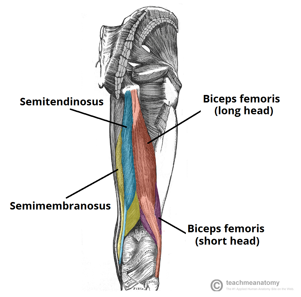
Muscles of the Posterior Thigh - Hamstrings - Damage ... from teachmeanatomy.info It is absent in 10% of people. Apr 23, 2019 · the plantaris is a small muscle with a long tendon, which can be mistaken for a nerve as it descends down the leg. The biceps includes a "short head" and a "long head" that work as a single muscle. Originating below and beneath the gastrocnemius is the soleus muscle, which extends your foot when your knee is bent. The muscle descends medially, condensing into a tendon that runs down the leg, between the gastrocnemius and soleus. The muscles tested, segmental level, and grading of dtr's are listed below. During an open surgery, an incision is made in the back of the leg and the achilles tendon is stitched together. The tendon continues its way through the foot by extending over its dorsal surface and finally inserting on the superior surface of the base of the distal phalanx of the hallux.
The tendon continues its way through the foot by extending over its dorsal surface and finally inserting on the superior surface of the base of the distal phalanx of the hallux.
The biceps is attached to the arm bones by. Originating below and beneath the gastrocnemius is the soleus muscle, which extends your foot when your knee is bent. The muscles tested, segmental level, and grading of dtr's are listed below. The muscle descends medially, condensing into a tendon that runs down the leg, between the gastrocnemius and soleus. In a complete or serious rupture the tendon of plantaris or another vestigial muscle is harvested and wrapped around the achilles tendon, increasing the strength of the repaired tendon. The posterior upper leg muscles provide your knees with mobility (extension, flexion and rotation) and strength.they work closely with your quadriceps muscles at the front of your thigh, your gluteal muscles, and your calf muscles to ensure proper movement of your leg and hip. The tendon continues its way through the foot by extending over its dorsal surface and finally inserting on the superior surface of the base of the distal phalanx of the hallux. During an open surgery, an incision is made in the back of the leg and the achilles tendon is stitched together. It is absent in 10% of people. It's the area that runs from the hip to the knee in each leg. It serves to attach the plantaris, gastrocnemius (calf) and soleus muscles to the calcaneus (heel) bone. The biceps is a muscle on the front part of the upper arm. Originates from the lateral supracondylar line of the femur.